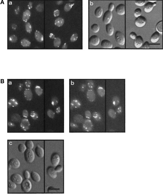
Figure 3.
Arc35-GFP colocalizes with filamentous actin. (A) A Wt strain expressing Arc35-GFP (RH4161) was visualized with epifluorescence (a) or Nomarski optics (b). Bar, 5 μm. (B) A Wt strain expressing Arc35-GFP (RH4161) was grown at 24°C, stained for F-actin with rhodamine–phalloidin, and visualized either with Nomarski optics (c) or with epifluorescence filters suitable to distinguish between GFP and rhodamine signals. The F-actin staining is shown in a, and the signal of Arc35-GFP is shown in b. Bar, 5 μm.

