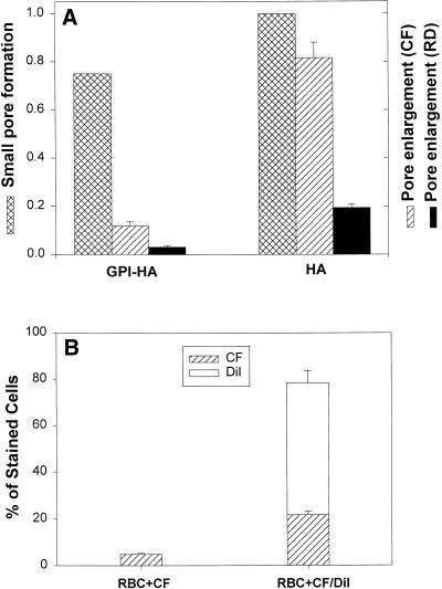
Figure 4.
Enlargement of fusion pores induced by GPI-HA and HA. (A) Fusion was triggered by locally applying pH 4.8 at 37°C for 2 min or until a pore formed. The fraction of bound RBC ghosts coloaded with CF and RD that fused was determined electrically (cross-hatched bars) by counting capacitative discharges resulting from fusion. In separate measurements of aqueous dye spread, the same batch of RBC ghosts was used and fusion was triggered at 37°C by reducing pH to 4.8 for 2 min followed by reneutralization at room temperature. Dye spread was monitored 10 min after acidification. Although, as determined electrically (cross-hatched bars), pores formed for almost the same percentage of GPI-HA cells with a bound RBC as for HA cells, CF (striped bars) did not pass as readily through GPI-HA pores. For HA cells, pores enlarged sufficiently to permit CF to transfer into 80% of the cells and to permit RD (solid bars) to transfer into ∼20% of the cells. (B) Transfer of CF through GPI-HA pores (striped bars) is greater when the RBC ghosts are labeled with DiI (second bar). But only ∼25% of the GPI-HA cells that became stained with DiI (open bar) also received CF. Different batches of cells were used in A and B; consequently, their extents of CF transfer are somewhat different.

