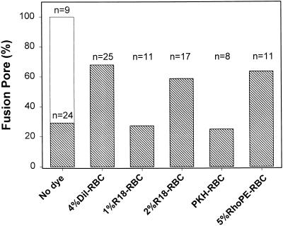
Figure 6.
The extent to which GPI-HA pore formation depends on the presence of lipid dye. RBCs, either unlabeled or labeled with the indicated membrane dye, were bound to GPI-HA–expressing cells, and a GPI-HA cell (with one bound RBC) was patch clamped in the whole-cell mode. Pore formation was detected by electrical admittance measurements, and the redistribution of a fluorescent dye was monitored simultaneously by fluorescence microscopy. Fusion was triggered at 30°C by applying a pH 4.8 solution. Either a pore formed or a pore did not form, but lipid dye always spread in virtually every experiment (i.e., every GPI-HA cell fused or hemifused). In the case of unlabeled RBCs, if a fusion pore did not form by 5 min after exposure to low pH, the experiment was terminated. In contrast to GPI-HA cells, pores always formed between HA cells and unlabeled RBCs (open bar) under these conditions. A higher percentage of fusion was generally observed with RBC ghosts (Figure 4, cross-hatched bars) than for intact RBCs (this figure). All experiments of this study, except for those shown in Figure 4, used intact RBCs. The numbers of experiments (n) carried out for each condition are given above the bars.

