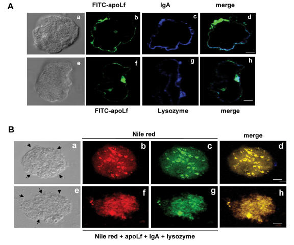
Figure 5.
Proteins of human milk bound to amoebic surface and damaged the trophozoites. (A) Amoebas (1x10⁵) were placed onto slides and then washed and incubated with FITC-apo-lactoferrin or unlabeled IgA and lysozyme (100 μM each). After 1 hour, samples were washed and incubated with anti-IgA or with anti-lysozyme for 1 hour, washed and incubated with secondary antibodies coupled to Cy5. (B) 1x10⁶ trophozoites were treated with Nile red for 30 minutes, washed and treated with IgA, apo-lactoferrin and lysozyme (1 mg/ml each) for 1 hour. All experiments were done at 37°C. Samples were processed to be analyzed by confocal microscopy. Arrows signal in (e) the damaged membranes, and photos are representative of the amoebic population. Bar, 8 μM.

