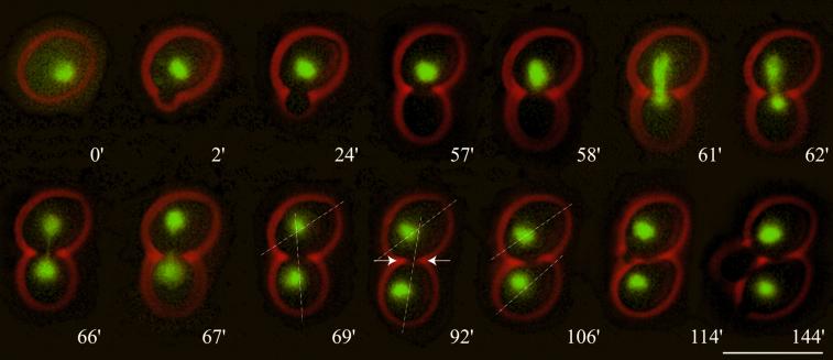
Figure 1.
Nuclear migration dynamics of diploid Hhf2-GFP–labeled wild-type cells observed by in vivo time-lapse microscopy. (0′) At onset of bud emergence the nucleus is positioned randomly. (2′–57′) During the positioning phase the nucleus moves to the bud neck region. (58′) The nucleus starts to elongate, marking the onset of anaphase B. (58′–62′) In a rapid process the nucleus becomes bar shaped, elongates along the mother–bud axis, and starts to penetrate the bud neck. This corresponds to spindle orientation, elongation, and penetration. (62′–67′) The labeled chromosomal material transforms into an hourglass shape. This phase is typically accompanied by bending and rapid oscillations of the nucleus within the bud neck. This can be seen by the fast change in position of the elongated nucleus. (69′) Final separation of chromosomal masses. At this time point the separated chromosomal masses show slow coordinated movements indicating that the spindle is still intact. (92′) Onset of cytokinesis can be observed by a slight change of the orientation of the longitudinal axis of the daughter cell relative to that of the mother and by a constriction of the bud neck (indicated with arrows). (106′) A sudden change of the orientation of the daughter cell relative to that of the mother indicates completion of cell separation. (114′) Next bud emergence of the mother cell. (144′) First distal bud emergence of the daughter cell. Bar, 10 μm. Movie 1: Nuclear dynamics as seen in Hhf2-GFP–labeled wild-type cells followed for >10 h. Two delayed mitoses can be seen in the second half of the time-lapse sequence. Acquisition interval, 1 min; movie speed, 10 frames per second = 10 min/s. One z-axis plane fluorescence image was acquired.

