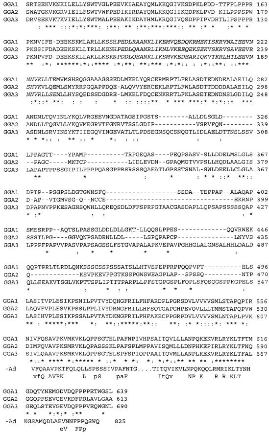
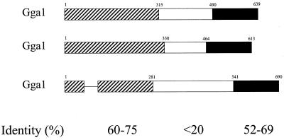
Figure 2.
Alignment of the protein sequences of GGA1, GGA2, and GGA3 showing regions of homology to γ-adaptin and predicted coiled-coil domains. (A) The amino acid sequences for GGA1, GGA2, and GGA3 were aligned with the use of CLUSTAL. Identical residues are indicated by asterisks (*) in the line below, and conservative substitutions are indicated by colons (:). Residues predicted to be in a coiled-coil configuration are italicized (begins at residue 188 for GGA1). The alignment with the C terminus of γ-adaptin (-Ad) is also shown, with the line below that showing identities in all four sequences (uppercase) or in three of the four sequences (lowercase). (B) The three domains with different levels of sequence conservation between GGAs are shown schematically. Bars representing the protein domains are scaled to reflect the relative size of each domain. The percent pairwise identity within each domain is indicated, as determined by Bestfit (Genetics Computer Group, Madison, WI).


