
Figure 2. ProMMP-2 is activated by RPE cells.
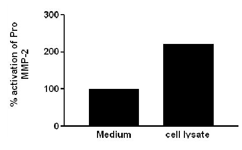
Graphic depiction of a two fold increase in pro MMP-2 activation in the presence of GFP-RPE cell lysates compared to medium alone. Ten μg of GFP-RPE cell lysates and 10mU of pro MMP-2 were incubated and analyzed by zymography. Data are expressed as percent activation of pro-MMP-2. Results are representative of two individual experiments performed in duplicate on cultured cells.