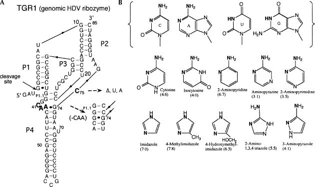
FIGURE 1.
(A) Sequence and secondary structure of the HDV genomic ribozyme and variants used in this study. The complete sequence of the wild-type ribozyme (TGR1) is shown. Changes at position 75 included deleting the nucleotide (C75Δ) or substituting with U or A (C75U, C75A). The sequence C41A42A43 was deleted from the C75Δ variant to generate the C75ΔC41Δ construct. (B) Structures of the four natural nucleobases and the exogenous bases used in the rescue reactions. The solution pKa values for the bases are given in parentheses.

