
Figure 2.
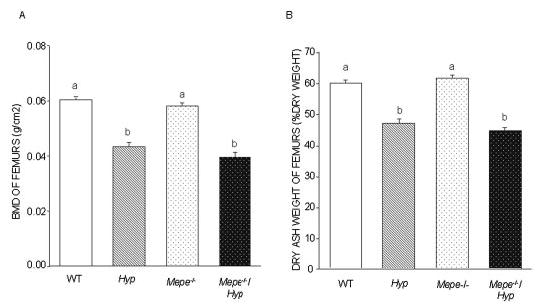
BMD and dry ash weight of femurs in WT and mutant mice. (A) BMD of the femur was assessed with the PIXImus mouse densitometer in 13-wk-old male and female WT, Hyp, Mepe−/−, and Mepe−/−/Hyp mice. (B) Dry ash weight was assessed in 13-wk-old male and female WT, Hyp, Mepe−/−, and Mepe−/−/Hyp mice. The decrease in mineralization of the skeleton in the Hyp compared with WT was confirmed by significantly reduced bone ash weight and BMD; however, Mepe−/−/Hyp exhibited no significant increment in bone ash weight and BMD compared with Hyp mice. Values sharing the same superscript are not significantly statistic different at P < 0.05.