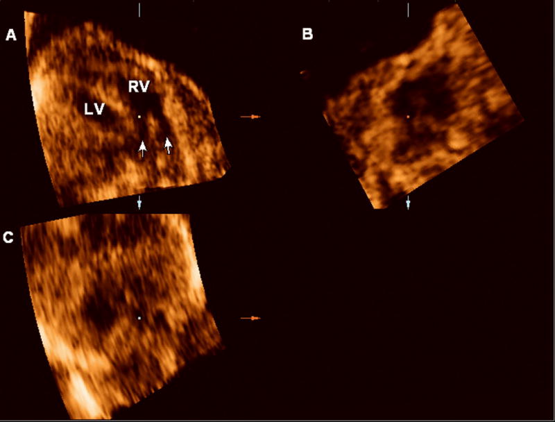
Figure 3.

4D multiplanar display of the fetal heart. Volume acquired with a transverse sweep through the fetal chest during excessive fetal motion. Panel A: Two vessels (arrows) apparently connect to the right ventricle (RV), suggesting a double outlet right ventricle. Panel B: sagittal view. Panel C: coronal view.
