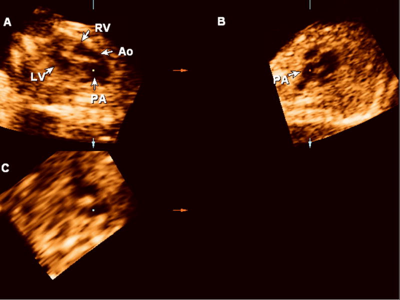
Figure 4.

Retrospective review of the volume dataset acquired using sagittal sweeps through the fetal chest reveals two vessels leaving the ventricles in parallel and the correct diagnosis of transposition of the great arteries (Panel A). Panel B: sagittal view. Panel C: coronal view. Ao: aorta; LV: left ventricle; PA: pulmonary artery.
