
FIGURE 1.
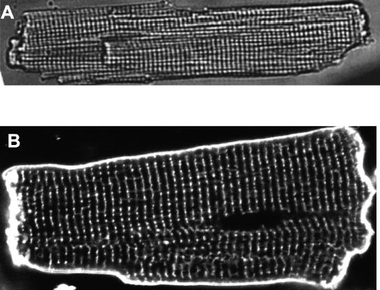
Transverse tubules (TTs) in rat heart cells. (A) A wide field transmitted light image of a rat ventricular myocyte. (B) A confocal fluorescence image of the TTs in a living rat ventricular myocyte stained with the membrane marker di-8 ANEPPS imaged with 488 nm excitation. The myocyte was exposed to 10 μM dye for 10 min. The striation or TT separation along the long axis of the cell is 1.8 μm.