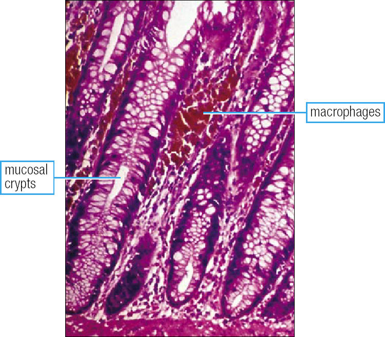
Figure 1.

The histologic appearance of melanosis coli, showing pigment-laden macrophages between the crypts in the lamina propria. The pigment is probably lipofuscin released by damaged epithelial cells. This is highly suggestive of long-term anthraquinone use. The effect is reversible within 1 year of discontinuing anthraquinone ingestion. The pigment-laden macrophages may be present histologically even when the endoscopic appearance of mucosa is normal. Hematoxylineosin stain, ×120. Reproduced with permission from reference 21.
