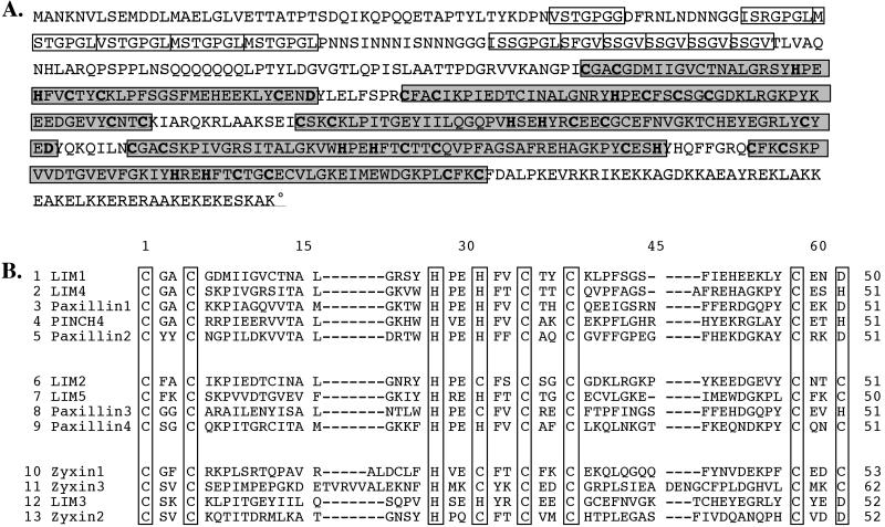
Figure 1.
Sequence and analysis of the domains of LIM2. A shows the derived amino acid sequence of LIM2. The five LIM domains are shaded. The NH2-terminal repeats are boxed, and the highly charged C-terminal stretch is underlined. B shows an alignment of the LIM domains from several Group 3 LIM domain-containing proteins. The domains are numbered with the most N-terminal domain numbered 1. The LIM domains of LIMB are aligned with all the LIM domains of chicken paxillin and the vertebrate Zyxin protein. The LIM domains of LIM2 show the strongest overall sequence homology to the LIM domains of paxillin, with the highest homology (57%) between the fourth LIM domain of LIM2 and the first LIM domain of paxillin. The PINCH LIM domain has 55% homology to the fourth LIM domain of LIM2. The other LIM domains of PINCH, however, do not show this high level of homology to any of the LIM2 LIM domains, despite similarities in the number and arrangement of the domains within these two proteins (our unpublished results).

