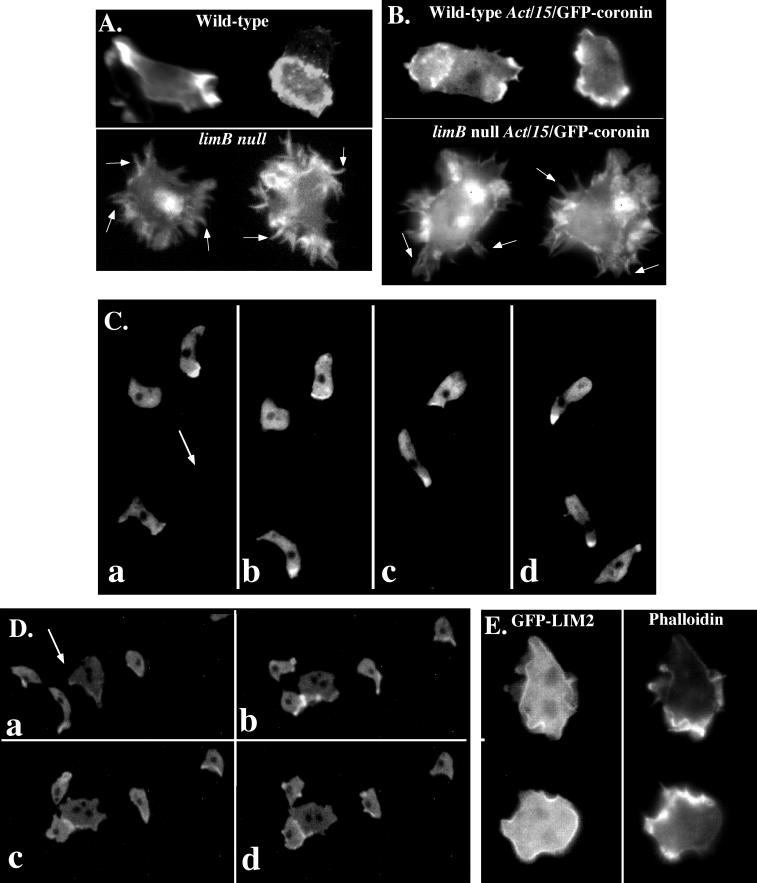
Figure 2.
F-actin cytoskeletal staining and subcellular localization of LIM2. (A) limB null and KAx-3 cells were fixed and labeled with FITC-conjugated phalloidin to show the actin cytoskeleton as previously described (Chung and Firtel, 1999). (B) limB null and KAx-3 cells expressing GFP–coronin, which binds to F-actin. In both A and B, the white arrows indicate actin rich “microspike” structures observed in the limB null cells. C and D show the localization of F-actin in wild-type and limB null aggregation streams using GFP–coronin. Wild-type (C) or limB null (D) vegetatively growing cells expressing GFP–coronin were washed and mixed with an ∼10-fold excess of unmarked wild-type cells and plated for development on thin agar. Once aggregation streams were formed, the cells were photographed using time-lapse video microscopy with either a Nikon (Melville, NY) Microphot FX or an Eclipse TE300 microscope with a 20× objective and equipped with fluorescence. The images were directly captured onto a computer using IP Labs software. Only the GFP–coronin–expressing cells can be seen. The direction of movement is indicated by the arrow. A series of sequential images are shown. E shows the subcellular localization of LIM2. LIM2–GFP fusion, which complements the null phenotype and was expressed from the Act15 promoter, is shown in the left panel. Phalloidin staining of F-actin is shown in the right panel.

