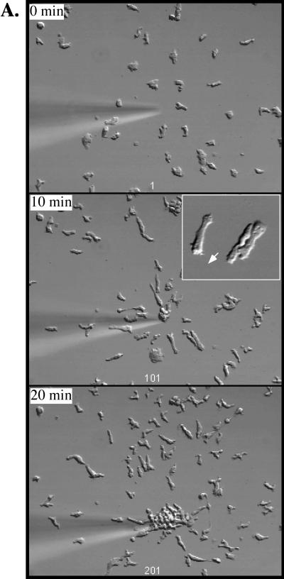
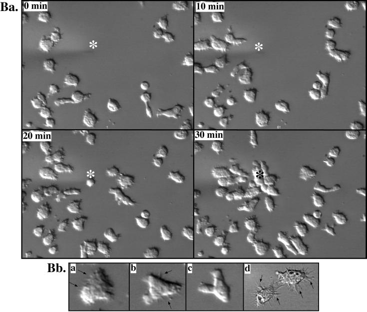
Figure 5.
Chemotaxis of cells to a micropipette containing the chemoattractant cAMP. Wild-type cells (A) or limB null cells (B) were washed and pulsed with 30 nM cAMP for 5 h to maximize expression of aggregation-stage chemotaxis signaling pathways (Insall et al., 1994; Ma et al., 1997; Meili et al., 1999). Cells were plated on a glass coverslip and allowed to adhere, and a micropipette containing cAMP was inserted (for details see Lee et al., 1999; Meili et al., 1999) (see MATERIALS AND METHODS). Insert in A shows an enlargement of wild-type cells. Ba shows the chemotaxis of limB null cells. Bb (a–c) shows an enlargement of three of these cells. Bb (d) shows a DIC image of two cells taken with a 60× objective. Arrows point to filopodia.


