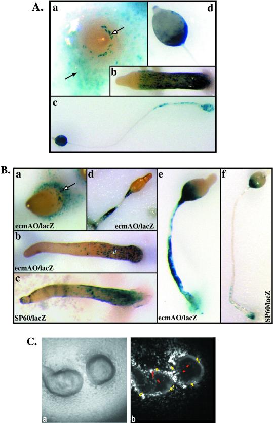
Figure 8.
Spatial localization of limB null cells in multicellular organisms. (A) One part limB null cells expressing Act15/lacZ were mixed with three parts KAx-3 cells. Aggregates were stained for β-galactosidase activity at various stages of development. (a) Mound stage. Labeled cells are primarily in the periphery of the mound (see open arrows). A few cells can be seen in the mound. (b) Slug stage. Stained cells are in a surface layer around the posterior 75% of the slug. (c) Fruiting body. Stained cells are found on the surface of the stalk and lower cup. (d) Closeup of the head of a fruiting body (sorus) showing staining of the lower cup or cells surrounding the lower portion of the sorus. (B) limB null cells expressing either ecmAO/lacZ or SP60/lacZ were mixed 1:3 with KAx-3 cells and stained for β-galactosidase activity at various developmental stages. (a) Tipped aggregate, limB null ecmAO/lacZ:KAx-3. (b) Slug, limB null ecmAO/lacZ:KAx-3. (c) Slug, limB null SP60/lacZ:KAx-3. (d) Early culminant, limB null ecmAO/lacZ:KAx-3. (e) Culminant, limB null ecmAO/lacZ:KAx-3. (f) Culminant, limB null SP60/lacZ:KAx-3. (C) One part limB null cells expressing Act15/GFP were mixed with 10 parts wild-type cells, and time-lapse movies were captured. (a) Bright-field view of two chimeric mounds. Movies indicate a rotational flow characteristic of wild-type cells of this strain. (b) Fluorescent image of the same mounds. Most of the limB null cells are found at the mound periphery, with a much smaller number of mutant cells located inside the mounds. Time-lapse movies of these mounds reveal that the limB null cells jiggle largely in place, either at the mound periphery or inside the mound. Cell trajectories are indicated by colored dots, yellow for cells at the periphery and red for cells in the mound interior. Each trajectory is composed of 25 dots representing cell position at 2-min time intervals over a period of 50 min. Because of the defective motion of the limB null cells, most of the dots are superimposed, and therefore the trajectories appear as large spots.

