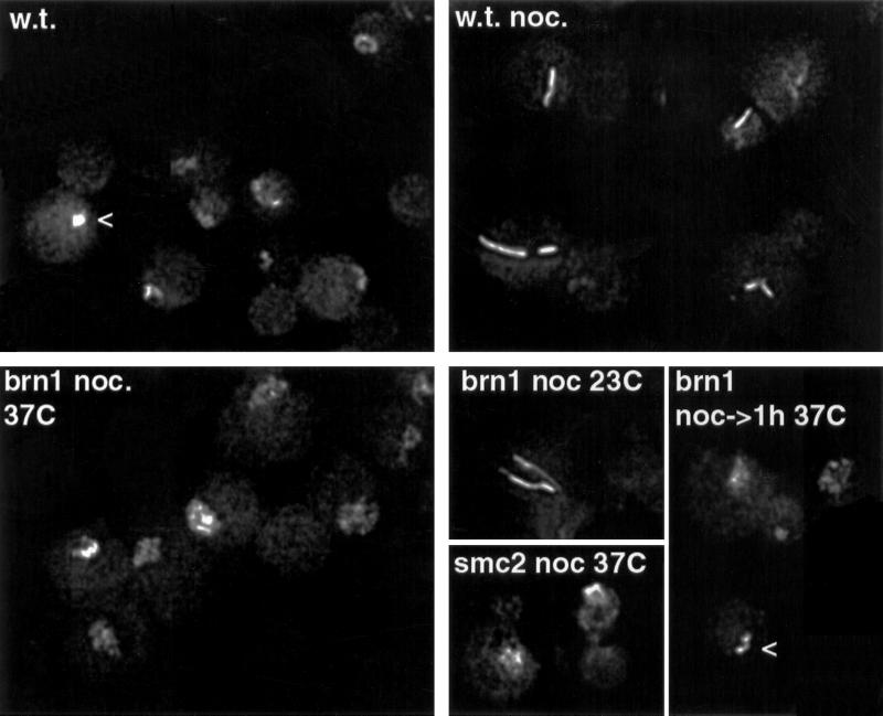
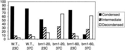
Figure 1.
Mitotic chromosome condensation defect in brn1 mutant cells. Examples of rDNA array morphologies, visualized by fluorescence in situ hybridization, are shown. BRN1 (w.t.) or brn1–60 cells were incubated at indicated temperatures for 3.5 h with or without nocodazole (noc). rDNA morphology in smc2–8 cells is shown for comparison. The bottom right panel (noc–>1h 37C) shows brn1–60 cells blocked in nocodazole at permissive temperature for 2.5 h, followed by a shift to 37°C in the continued presence of nocodazole. Arrowheads point to examples of rDNA morphology scored as “intermediate.” The graph below shows percentages of cells with the indicated rDNA morphologies after 3.5 h at the restrictive temperature in the presence of nocodazole. Blind scoring of at least 100 cells in each preparation was performed. Cells that could not be unequivocally assigned to one of the two classes were scored as intermediate (see arrowheads above).


