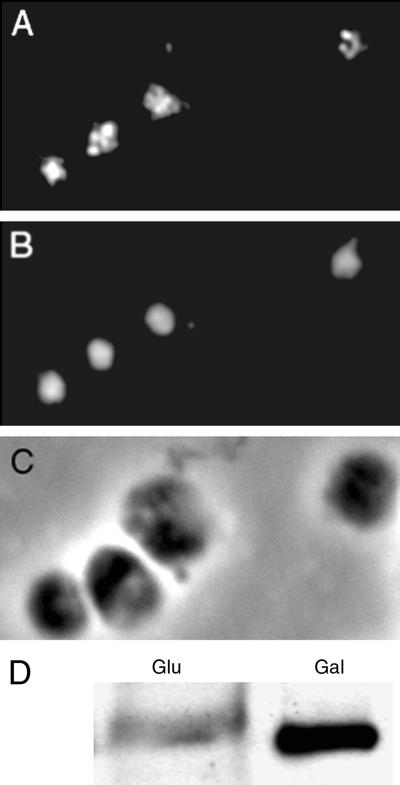
Figure 6.
Intracellular localization of the Brn1p protein. (A) Immunofluorescent staining of cells overexpressing Brn1p from the GAL1 promoter on a CEN plasmid, using an affinity-purified anti-Brn1p antibody. (B) Nuclear DNA of the same cells stained with DAPI. (C) Phase-contrast image of the same cells. (D) Levels of Brn1p under induced (Gal) and uninduced (Glu) conditions.

