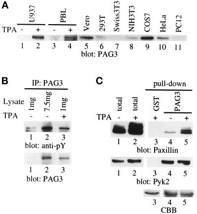
Figure 2.
Induced expression of PAG3 and its increased binding to paxillin during monocyte maturation. (A) Cell lysates from U937 monocytes, human peripheral monocytes (PBL), and several other cell lines were separated on SDS-PAGE and subjected to immunoblot analysis using anti-PAG3 antibody. Monocyte cells undifferentiated (−TPA) or differentiated by treatment with TPA for three days (+TPA) are shown. (B) PAG3 protein was immunoprecipitated from U937 cells treated with (lane 3) or without (lanes 1 and 2) TPA and subjected to sequential immunoblot analysis using anti-phosphotyrosine antibody (4G10; anti-pY) and anti-PAG3 antibody. Different amounts of cell lysates (1 mg in lane 1, 7.5 mg in lane 2, and 1 mg in lane 3) were initially used for comparison of the tyrosine phsosphorylation levels. (C) Cell lysates from TPA-treated or untreated U937 cells were incubated with GST-PAG3 purified on glutathione beads to analyze PAG3 binding toward paxillin and Pyk2. Immunoblots were done with same membrane filter by sequential hybridization with anti-paxillin antibody (Ab 199–217, Pax) and anti-Pyk2 antibody (Pyk2). In C, amounts of each fusion protein used for pull-down assays (lane 3, GST; lanes 4 and 5, GST-PAG3) are shown by Coomassie brilliant blue staining (CBB).

