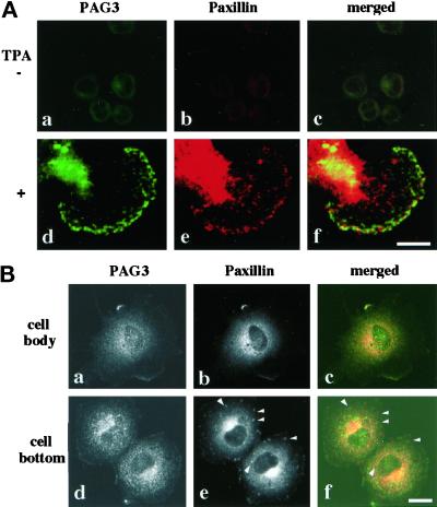
Figure 3.
Colocalization of endogenous PAG3 and paxillin in the cytoplasm and at the cell periphery in U937 monocyte cells (A) and COS-7 epithelial cells (B). U937 monocyte cells undifferentiated (A, a–c) or differentiated by TPA treatment for 3 d (A, d–f) are shown. Cells fixed with 3.7% paraformaldehyde were double immunolabeled with rabbit polyclonal anti-PAG3 antibody (a and d) and mouse monoclonal anti-paxillin antibody (b and e), followed by Cy2-conjugated donkey anti-rabbit IgG and Cy5-conjugated donkey anti-mouse IgG, and examined by confocal laser scanning microscope. Focuses were adjusted 5.0 μm above the surface of the glass chamber plate in A (a–c) and 3.0 μm above this in B (a–c), each across the center of the nucleus in the majority of the cells, or 0.5 μm above this in A (d–f) and B (d–f), which are near the bottom layers of cells. In e and f in B, paxillin localized at the cytoplasmic pool is still seen in addition to that localized to focal adhesion plaques, and several focal adhesion plaques are marked by arrowheads. The right column represents the merging of the left and the middle images. For clearer images of PAG3 and paxillin distribution, some photographs in B (a, b, d, and e) are shown as black-and-white images. Bars, 20 μm.

