Figure 5.
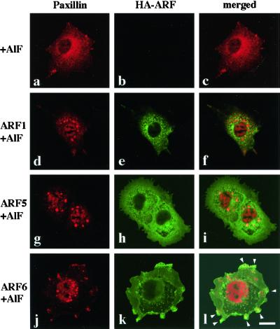
ARF activities affect subcellular localization of paxillin. COS-7 cells untransfected (a–c) or transiently transfected by FuGENE 6 with each plasmid encoding HA-tagged wild-type ARF1 (d–f), ARF5 (g–i), or ARF6 (j and k) were treated with AlF for 1 h at 37°C. Cells were then fixed, and endogenous paxillin (a, d, g, and j) was visualized using rabbit polyclonal anti-paxillin antibodies, coupled with Cy5-conjugated donkey anti-rabbit antibody, and HA-ARF proteins (e, h, and k) were visualized mouse monoclonal anti-HA antibody, coupled with Cy2-conjugated donkey anti-mouse antibody. Focuses were adjusted 3.0 μm above the surface of the glass chamber plate. The right column represents the merging of the left and the middle images (c, f, i, and l). Arrowheads in panel l indicate areas where paxillin is colocalized with ARF6. Bar, 20 μm. Expression of these wild-type HA-ARF proteins in the absence of the AlF treatment did not affect significantly the subcellular localization of paxillin (our unpublished data): see Figure 3B (b) for the subcellular distribution of paxillin in AlF-untreated cell for the comparison.

