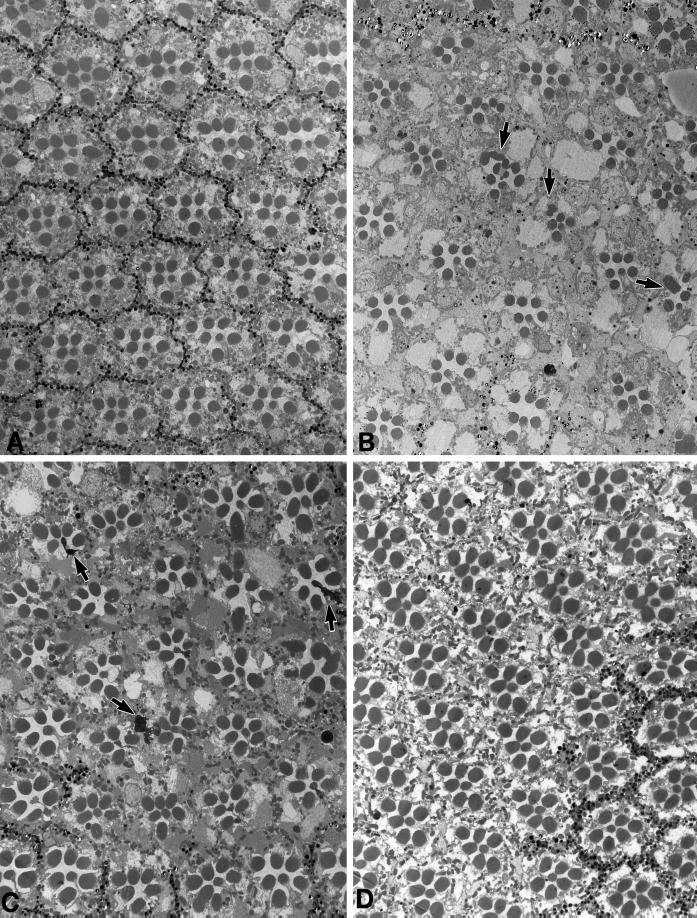
Figure 5.
Effects of a loss of KHC on photoreceptor cells. (A) Transmission electron micrograph of a cross section from a wild-type retina. Ommatidia normally have eight photoreceptor cells, each containing a photosensitive rhabdomere. The rhabdomeres of R1–6 are located around the outside of the ommatidium and extend along its entire length (∼100 μm). The rhabdomeres of R7 and R8 are located in the center with R7 stacked above R8. Thus, seven rhabdomere profiles are visible in most cross sections. (B) Portion of Khcnull eye clone from a newly eclosed fly. Khcnull ommatidia can be distinguished by the absence of the pigment granules that normally surround them. Note that a few null ommaditia have abnormal or missing photoreceptor cells (arrows). (C) Portion of a Khcnull eye clone from a fly aged 3 wk after eclosion. In addition to the structural defects seen in the photoreceptors of null clones in newly eclosed flies, some photoreceptors have degenerated (arrows). (D) Portion of a control Khcnull eye clone from a fly aged 3 wk after eclosion. The transgenic copy of wild-type Khc rescued the developmental defects as well as the age-dependent photoreceptor degeneration.

