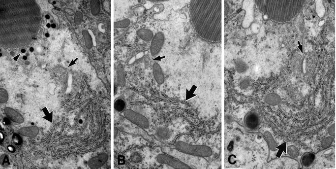
Figure 6.
ER and Golgi organization in Khcnull photoreceptor cells. The three photoreceptor cells pictured here were from adjacent ommatidia located on the border of an eye clone that contained >1400 Khcnull cells. (A) Cross section of a wild-type photoreceptor cell. (B and C) Equivalent cross sections of null photoreceptor cells. Arrows indicate rough ER (large arrows) and Golgi bodies (small arrows). The genotypes of all cells examined were assessed by the presence (wild-type; Khc27/+ or +/+) or absence (null: Khc27/Khc27) of ommachrome pigment granules (A, arrowhead) adjacent to the base of the rhabdomere. Golgi morphology and ER organization in null and wild-type photoreceptors showed no consistent difference.

