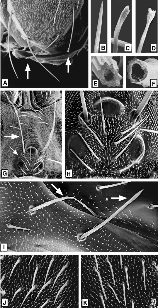
Figure 7.
Effects of a loss of KHC on secreted cuticle structures. Mitotic recombination was used to generate Khc20/Khc20 null clones marked with yellow bristles as for Figure 3. Clones were mapped by light microscopy and then examined in detail by SEM. (A) Scutellum with a large null clone covering the right half. Two null scutellar bristles (∼400 μm long) are marked by arrows. (B–D) High-magnification views of the tips of three of the scutellar bristles shown in A. (B) Wild type, (C and D) null. (E and F) Bases of wild-type (E) and Khcnull (F) scutellar bristle shafts that were broken off near their sockets. (G) Dorsal side of a head containing a Khcnull clone. The single macrochaete (∼280 μm long; large arrow) and the four microchaetae (small arrow) on the left side were null, whereas the analogous bristles on the right side were wild type. (H) A higher-magnification view shows that the null microchaetae (∼80 μm long) have slight tip abnormalities. (I) Mutant abdominal macrochaetae (∼170 μm long; white arrows). Note that the null sockets and surrounding sheet of cuticle appear normal here and in the other micrographs. (J) High-magnification view of wild-type microchaetae (∼70 μm long) and epidermal hairs (∼10 μm long) from the left side of a thorax. (K) Analogous area on the right side of the same thorax in the center of a large null clone. Note that mutant microchaetae in the clone have aberrant tips, whereas there are no apparent defects in the epidermal hairs.

