Figure 1.
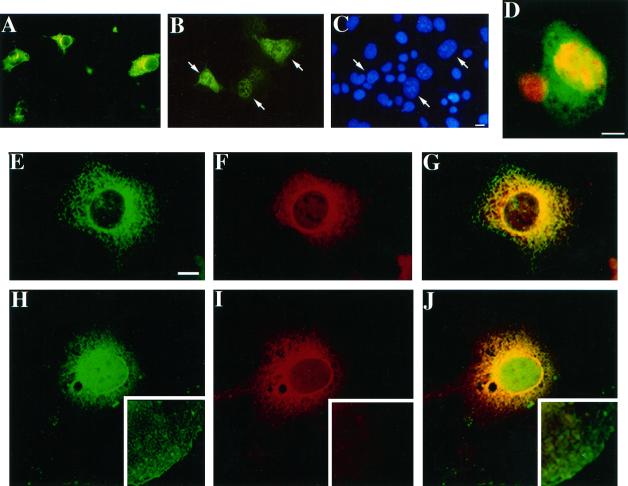
Cellular localization of secreted and iLIF proteins in Cos-1 cells transfected with LIF expression vectors 2 d after transfection. (A) LIF-D (pmLIF-DX)-transfected cells stained with anti-mLIF. (B) LIF-T (pmLIF-TX)-transfected cells stained with anti-mLIF. (C) Cells in B stained with Hoechst DNA stain. Cells were visualized using conventional optics. Arrows indicate selected LIF-staining cells and their nuclei. (D) Confocal laser scanning image of a Cos-1 cell transfected with an expression vector for LIF-T (pmLIF-TX) stained for LIF protein (green) and DNA (red). Colocalization of LIF protein and DNA in the nucleus appears yellow. The red nucleus (lower left) is an untransfected cell. (E–J) Confocal laser scanning images of Cos-1 cells transfected with pmLIF-DX (E–G) and pmLIF-TX (H–J) stained with anti-LIF (E and H) and concanavalin A (F and I). Merged images are presented in G and J. Insets show contrast-enhanced regions of the corresponding cellular cytoplasm. Bars, 10 μm.

