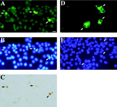
Figure 2.
Overexpression of iLIF but not secreted protein induces apoptosis. Cos-1 cells transfected with the LIF-T expression vector pmLIF-TX were stained 3 d after transfection for LIF protein (A), DNA (B), and internucleosomal DNA fragmentation (C). Arrows indicate the nuclei of selected LIF-staining cells. Apoptotic cells with varying degrees of DNA condensation and fragmentation are shown. The plane of focus in B is that of the rounded apoptotic cells, seen most clearly in the central cell. Elevated background staining in A reflects interference from antibodies used in costaining for DNA fragmentation (Apoptag). Cos-1 cells transfected with pmLIF-DX stained with anti-LIF antibody (D) and Hoechst DNA stain (E) 3 d after transfection. The healthy nuclear morphology of pmLIF-DX-transfected cells is indicated by arrows. Bar, 20 μm.

