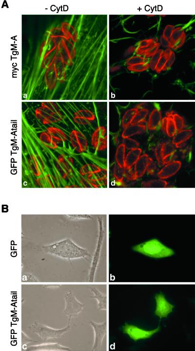
Figure 8.
Cytochalasin treatment did not influence the peripheral localization of mycTgM-A and GFP·TgM-Atail. GFP·TgM-Atail did not localize to the plasma membrane of HeLa cells. (A) MycTgM-A (a and b) and GFP·TgM-Atail (c and d) localized at the periphery of parasites after incubation in the presence (b and d) or absence (a and c) of 10 μg/ml cytochalasin D for 3 h, indicating that it did not depend on an intact actin cytoskeleton. To optimally present the host actin cytoskeleton and the peripheral staining in the parasites, one confocal section was recorded in the ventral part of the fibroblasts where the actin stress fibers are most prominent, and another confocal section was recorded through the middle of the parasites, 1 μm higher in the cell. Only the overlay is shown here. (B) Contrary to what was seen in T. gondii, GFP·TgM-Atail did not interact with the plasma membrane of an animal cell (d) and had a distribution similar to GFP (b). (a and c) Phase-contrast pictures corresponding to b and d.

