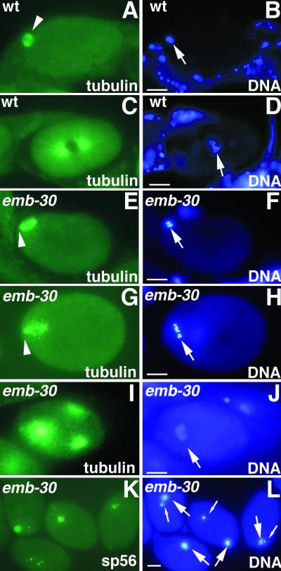
Figure 2.
emb-30 is required for completion of the oocyte meiotic divisions. Cytological analysis of embryos stained for tubulin, a sperm membrane protein (sp56), or DNA, as indicated. Wild-type (A and B) and emb-30(tn377ts) (E and F) embryos in MI metaphase. The MI spindle (arrowheads) is associated with the anterior cortex. (C and D) Wild-type pronuclear-stage embryo in prometaphase of the first mitotic division. (G and H) emb-30(tn377ts) one-cell embryo with a disorganized meiotic spindle (arrowhead). Six bivalents are visible (arrow), but there is no evidence of chromosome segregation. The spindle and chromatin are not closely associated with the anterior cortex. This particular spindle and chromatin morphology is not observed in the wild type. (I and J) emb-30(tn377ts) one-cell embryo with multipolar spindles. Three microtubule-organizing centers are seen. (K and L) emb-30(tn377ts) one-cell embryos stain for a sperm membrane protein and contain both maternal DNA (large arrows) and paternal DNA (small arrows). It is unclear how the female and male chromatin can on occasion come to be adjacent in the arrested embryos. This does not reflect nuclear migration processes because pronuclei do not form. Bars, 10 μm.

