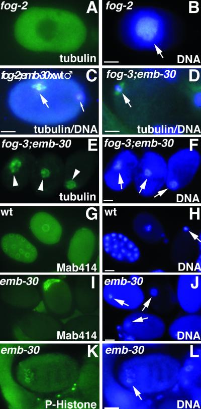
Figure 3.
Cytological analysis of embryos or oocytes stained for tubulin, nuclear pore complex (Mab414), phosphohistone H3 (P-Histone), or DNA, as indicated. (A and B) Unfertilized oocyte from a fog-2 female showing intense DAPI staining (arrow) and diffuse tubulin staining. (C) One-cell embryo after fertilization of a fog-2; emb-30(tn377ts) oocyte with wild-type sperm. The MI spindle (large arrow) is in metaphase but is not associated with the anterior cortex. The paternal DNA is in the posterior (small arrow) but slightly out of the plane of focus. (D) Unfertilized oocyte from a fog-3; emb-30(tn377ts) female. An MI spindle can assemble in the absence of fertilization (arrow) and associate with the anterior cortex, but the oocyte does not become endomitotic. (E and F) Unfertilized oocytes from fog-3; emb-30(tn377ts) females. Disorganized meiotic spindle–like structures (arrowheads) associate with the maternal chromatin (arrows). Note that there are three separate oocytes in panels E and F. (G and H) Wild-type embryos form nuclear envelopes. Gastrula-stage embryo (left), pronuclear-stage embryo (middle), and meiotic embryo (right). (I and J) In emb-30(tn377ts), there is no indication of nuclear envelope formation. Instead, an amorphous aggregate of nuclear pore complex material is found in 8% of the arrested embryos. Maternal DNA is indicated by arrows. (K and L) Less condensed chromatin (arrow) contains phosphohistone H3, indicating that the embryo arrests in M phase. Bars, 10 μm.

