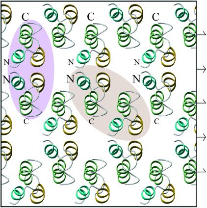
Figure 7.
Cooperative model of Er-1–Er-1mem interactions. The model is modified from the study of the Er-1 crystal structure by Weiss et al. (1995), and the representation was prepared by using the MOLMOL program (Koradi et al., 1996). On the basis of the structural similarity of the extracellular domain of Er-1mem with Er-1, it is assumed that Er-1–Er-1mem interactions on the cell surface mimic Er-1–Er-1 interactions in the crystal lattice. Half of the molecules are top viewed, with their N termini in the back and C termini in the front; they represent Er-1mem molecules on the cell surface. The other half are bottom viewed, with their N termini in the front and C termini in the back; they represent soluble Er-1 molecules. Back and front positions are indicated by smaller and larger letters, respectively. The two types of dimeric associations that each molecule can form with its neighboring molecules are outlined by ovals of different colors. Positions of the twofold rotation axes between molecules in dimers 1 and the twofold screw axes between molecules in dimers 2 are indicated on the margin by symmetrical and asymmetrical arrows, respectively.

