Figure 4.
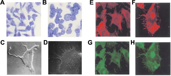
Analysis of the role of ADAM 23 in neuroblastoma cell adhesion. Adhesion of NB 100 cells to dishes coated with 10 μg/ml fibronectin (A) or with 10 μg/ml recombinant ADAM 23 (B) is shown. Several differences in cell morphology are found at the light microscopic level. Scanning electron microscopy proved that differences in cell morphology between NB 100 cells adherent to fibronectin (C) and those adherent to ADAM 23 (D) are mainly due to variations in the number and length of surface protrusions. Cells adherent to fibronectin (C) have a relatively low number of long asymmetric filopodial extensions, whereas those attached to ADAM 23 are devoid of long surface extensions but show many short protrusions resembling microspikes, most of which appeared to be firmly attached to the glass coverslip. The effect of ADAM 23 on F-actin and vinculin distribution is shown in E–H. Confocal optical sections of NB100 cells adherent to fibronectin (E and G) and ADAM 23 (F and H) were double labeled for F-actin (rhodamine-phalloidine; red) and vinculin (anti-vinculin, visualized with FITC-labeled goat anti-rabbit; green). Neuroblastoma cells adherent to fibronectin (E) show relatively little F-actin in the central region of the cell and numerous parallel bundles at the axis of the filopodial protrusions. In cells adherent to ADAM 23 (F), the assembly of actin filaments is lower, but they are mainly located at specific cortical regions and display a moderate polarization. Vinculin in neuroblastoma cells adherent to fibronectin (G) is mainly located at the sites of filopodial protrusion. Vinculin-positive patches in cells adherent to ADAM 23 (H) show a lower degree of aggregation, but they are also concentrated at specific cortical regions.

