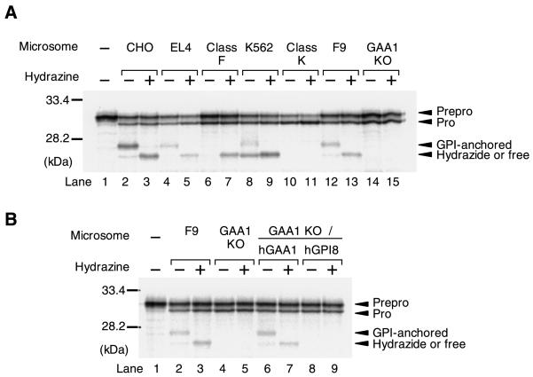
Figure 3.
Microsomal membranes from GAA1-knockout cells do not form a carbonyl intermediate of mini-PLAP. Capped mini-PLAP mRNA was translated in vitro using rabbit reticulocyte lysate and microsomal membranes prepared from the indicated cells in the absence (− or presence (+) of hydrazine. Lane 1 in both A and B shows a translation product in the absence of microsomal membranes, yielding prepro–mini-PLAP. Identities of other bands determined according to Kodukula et al. (1991) are shown on the right. Positions of prestained molecular markers are shown on the left. The cellular origin of the microsomal membranes is indicated as well as the presence or absence of hydrazine. In B, lanes 6 and 7 show the microsomal membranes from GAA1-knockout cells rescued with human GAA1 cDNA, and lanes 8 and 9 show those from GAA1-knockout cells transfected with human GPI8 cDNA.

