Figure 4.
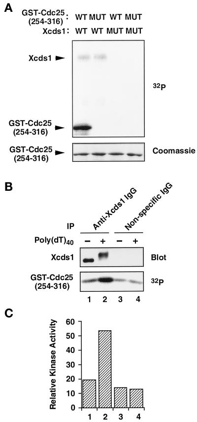
Activation of Xcds1 kinase in response to poly(dT)40. (A) His6-Xcds1 (WT) and His6-Xcds1-N324A (MUT) were purified from E. coli and incubated with GST-Cdc25[254–316]-WT (WT) or GST-Cdc25[254–316]-S287A (MUT) in kinase buffer containing [32P]ATP. The proteins were separated by SDS-PAGE and stained with Coomassie blue. The phosphorylated proteins were detected with the use of a PhosphorImager. (B) Immunoprecipitation (IP) was performed from interphase egg cytosol containing poly(dT)40 or no DNA with the use of anti-Xcds1 antibodies or nonspecific rabbit IgG. One-third of each immunoprecipitate was analyzed for the modification of Xcds1 by immunoblotting; two-thirds of each immunoprecipitate was incubated with GST-Cdc25[254–316]-WT to measure kinase activity as in A. (C) Quantitation of the data presented in B with the use of a PhosphorImager.

