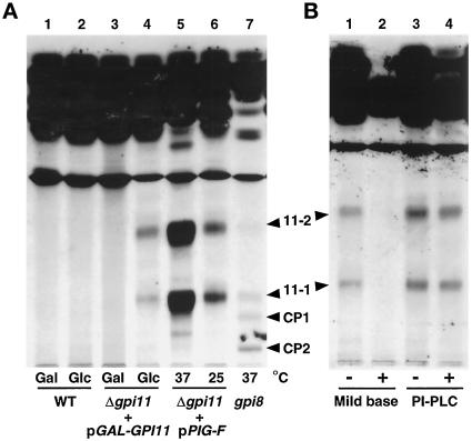
Figure 4.
Accumulation of mild base-sensitive and PI-PLC-resistant [3H]inositol-labeled lipids in gpi11:: LEU2-pGAL-GPI11 and gpi11:: LEU2-pPIG-F cells. (A) Wild-type (WT) and gpi11:: LEU2-pGAL-GPI11 (Δgpi11 + pGAL-GPI11) cells were maintained on SGlyYE medium, shifted to SGalYE or SGlcYE medium for 16 h, then resuspended in 1 ml of inositol-free medium containing Gal (lanes 1 and 3) or Glc (lanes 2 and 4), respectively, and radiolabeled with 15 μCi of [3H]inositol for 60 min at 30°C. gpi11:: LEU2-pPIG-F cultures were grown at 25°C in inositol-free medium and then divided into two equal portions, one of which was shifted to 37°C for 20 min and the other maintained at 25°C. Shifted and control cultures were then radiolabeled with 15 μCi of [3H]inositol for 2 h at 37 and 25°C, respectively (lanes 5 and 6). The gpi8 strain was grown in inositol-free medium, shifted to 37°C, and pulse labeled at 37°C (lane 7). Radiolabeled lipids were then extracted as detailed in MATERIALS AND METHODS. (B) Lipids from gpi11::LEU2-pGAL-GPI11 cells that had been shifted to glucose and [3H]inositol-labeled in glucose-containing medium were incubated with methanol/water or methanolic ammonia (mild base) (lanes 1 and 2) or without or with PI-PLC (lanes 3 and 4). After either treatment, incubation mixtures were extracted with buffer-saturated 1-butanol and submitted to phase partitioning, after which the butanol phase was separated by TLC using solvent B, and radiolabeled lipids were detected by fluorography. Arrowheads indicate the positions of lipids 11-1, 11-2, and complete precursors CP1 and CP2 (Sipos et al., 1994).

