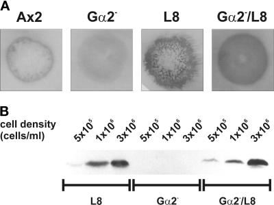
Figure 8.
Gα2−/gdt1− cells express high levels of discoidin. (A) Colony blot of Ax2, Gα2−, gdt1−, and Gα2−/gdt1− cells. Colonies of Ax2, Gα2−, L8, and Gα2−/L8 cells were stained with the anti-discoidin antibody. The colony blot shows the outer ring staining in Ax2, weak background staining in Gα2−, a strong label in L8, and also a strong signal in Gα2−/L8 double mutant cells. Aggregation and the irregular colony shape are not detected in the double mutant. (B) Western blot of Ax2, Gα2−, gdt1−, and Gα2−/gdt1− cells. For Western blot analysis, the double mutant and both single mutants were grown to the cell densities indicated, cells were harvested, and total protein was subjected to electrophoresis, blotted, and stained with the anti-discoidin antibody. The blot demonstrates that discoidin levels in the double mutant are similar to those in L8 cells and that discoidin accumulates with increasing cell density.

