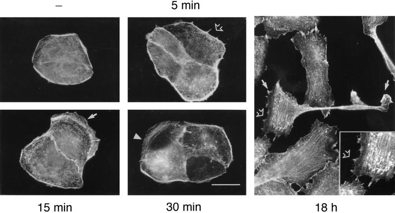
Figure 1.
HGF-induced actin reorganization in MDCK cells. MDCK cells were stimulated with HGF (5 U/ml) for the indicated times and fixed in 3.7% formaldehyde in PBS. After fixation, cells were permeabilized with Triton X-100 (0.2%), and filamentous actin was stained with TRITC-phalloidin. Note the appearance of microspikes at 5 min (open arrow) and lamellipodia at 15 min (thin arrow) after HGF stimulation. Membrane ruffles are found at the edge of lamellipodia (thick arrow), whereas by 30 min, actin fibers are present in the lamellipodia (arrowhead). After overnight stimulation (18 h), cells harbor filopodia-like structures (inset), lamellipodia, and membrane ruffles typical of motile cells. Bar, 25 μm.

