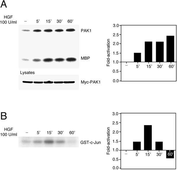
Figure 3.
HGF-induced PAK1 and JNK activation in MDCK cells. (A) MDCK cells stably expressing Myc-tagged PAK1 were serum starved for 24 h and then stimulated with HGF (100 U/ml) for the indicated times. PAK1 was immunoprecipitated from cell lysates with the Myc 9E10 mAb, and its kinase activity was assayed in the presence of the exogenous substrate MBP and [γ-32P]ATP. The reaction was stopped by adding Laemmli sample buffer, and proteins were separated by electrophoresis on a 15% SDS-polyacrylamide gel. Cell lysates were subjected to Western blotting with the 9E10 Myc mAb to show amounts of PAK1 present. (B) JNK activation was assayed in the presence of 10 μg of cell lysates, GST-c-Jun (aa 5–89), and [γ-32P]ATP, and proteins were resolved by SDS-PAGE (10%). The bar graphs indicate the activation of PAK1 and JNK detected over basal levels present in unstimulated cells.

