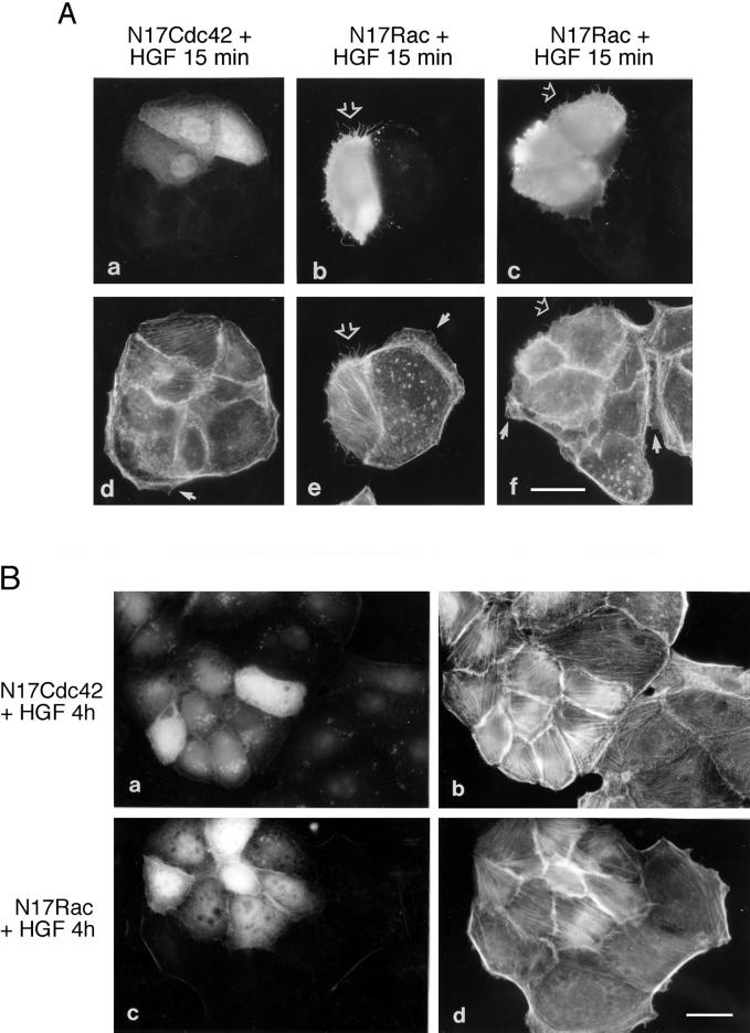
Figure 4.
Cdc42 and Rac are required for HGF-induced lamellipodia formation and cell spreading/dissociation. (A) cDNAs of N17Cdc42 and N17Rac (in pRK5Myc vector; 0.1 mg/ml) were injected into the nuclei of MDCK cells. After a 2-h incubation period, cells were stimulated with HGF (5 U/ml) for 15 min. Cells were fixed as described in Figure 1 and double stained with anti-rabbit FITC to identify the cells coinjected with rabbit immunoglobulin G (a) or with anti-Myc and anti-mouse FITC to detect protein expression (b and c), followed by TRITC-phalloidin (d–f) to stain actin. Note the presence of lamellipodia (arrows) and microspikes (open arrows). Bar, 25 μm. (B) MDCK cells were coinjected with N17Cdc42 (a and b) or N17Rac (c and d) together with rabbit immunoglobulin G. After a 2-h incubation period, cells were stimulated with HGF for 4 h, fixed as described, and double stained with anti-rabbit FITC (a and c) and TRITC-phalloidin (b and d). Bar, 25 μm.

