Figure 5.
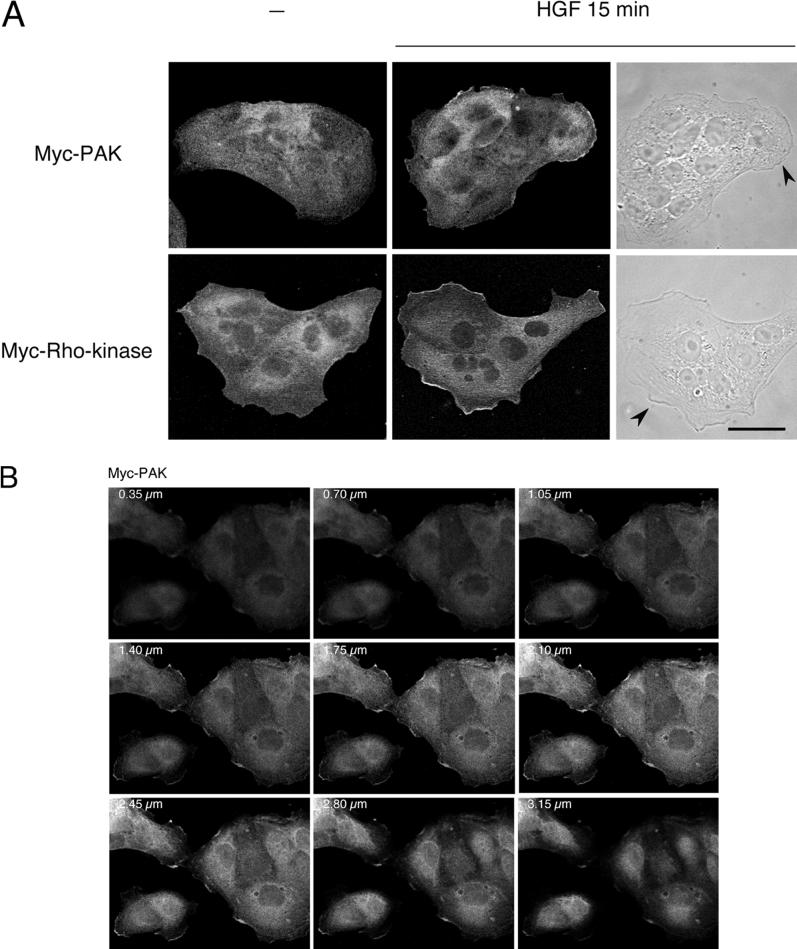
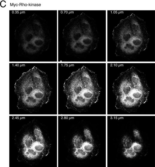
HGF stimulation induces membrane translocation of PAK1 and Rho-kinase. (A) MDCK cells stably expressing Myc-tagged PAK1 or Myc-tagged Rho-kinase were stimulated with HGF (5 U/ml) for 15 min. Cells were fixed in 3.7% formaldehyde in PBS, permeabilized in Triton X-100 (0.2%), and stained with anti-Myc and anti-mouse FITC. Fluorescent images were obtained by laser confocal microscopy. Note the presence of PAK1 and Rho-kinase in the membrane ruffles found at the edges of lamellipodia (arrowheads). Bar, 25 μm. (B and C) Confocal sectioning in the z-axis of cells depicting successive sections through the lamellipodia and membrane ruffles of HGF-stimulated cells expressing Myc-PAK and Myc-Rho-kinase.


