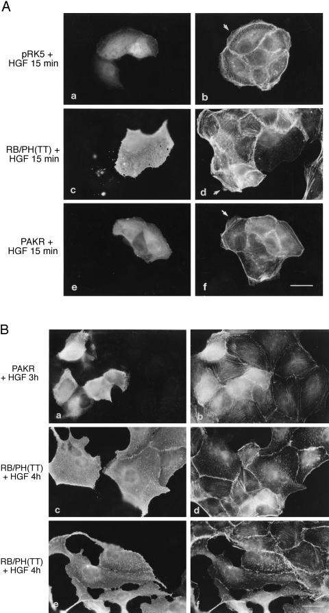
Figure 6.
Rho-kinase and PAK are involved in HGF-induced lamellipodia formation, but only PAK is required for MDCK cell spreading. (A) pRK5Myc vector (0.2 mg/ml) together with rabbit immunoglobulin G (a and b) or expression vectors (0.2 mg/ml) encoding Myc-tagged RB/PH(TT) (c and d) or Myc-tagged PAKR (e and f) were injected into the nuclei of MDCK cells. After a 5-h incubation period, cells were stimulated with HGF (5 U/ml) for 15 min. Cells were double stained with anti-rabbit FITC (a) and TRITC-phalloidin (b) or anti-Myc and anti-mouse FITC (c and e) and TRITC-phalloidin (d and f). Arrows point to lamellipodia. Bar, 25 μm. (B) MDCK cells were injected with expression vectors (0.2 mg/ml) encoding Myc-tagged PAKR (a and b) or Myc-tagged RB/PH(TT) (c–f). After a 2-h incubation period, cells were stimulated with HGF for 3 h (a and b) or 4 h (c–f) and then fixed and double stained with anti-Myc and anti-mouse FITC (a, c, and e) and TRITC-phalloidin (b, d, and f). Bar, 25 μm.

