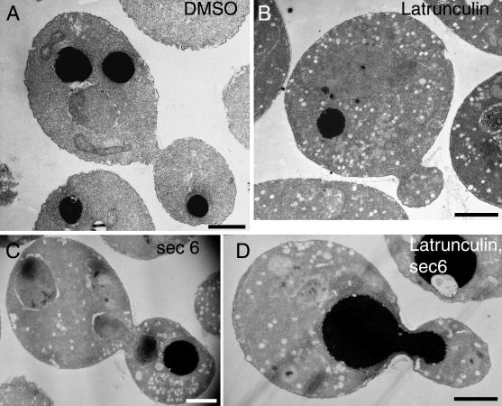
Figure 2.
Accumulation of vesicles in latrunculin-treated cells observed by thin-section electron microscopy. Wild-type cells (NY13) were grown at 25°C for 120 min in the presence of DMSO (A) or 200 μM latrunculin (B). sec6-4 cells (NY17) were grown at 37°C for 30 min in the presence of DMSO (C) or 200 μM latrunculin (D). Bars, 1 μm.

