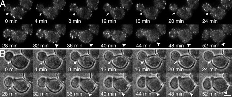
Figure 4.
Bud formation and cell separation in the absence of patch polarization in the bee1Δ(las17Δ) mutant (YJC1691). Observations were made at 4-min intervals over a 2-h period spanning the whole cell cycle. Sequential frames show fluorescence (A) and bright-field (B) images of the same cell. The arrows point to the site of cell separation, and the arrowheads point to the site of bud formation. Cell separation is indicated by a slight change in the position of the bud relative to the mother between 24 and 28 min. This movement is more obvious in the video sequence available online. Cortical actin patches were tagged with Cap1-GFP. Bar, 2.5 μm.

