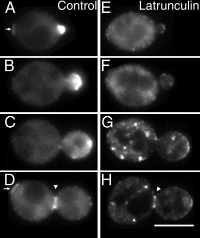
Figure 5.
Myo2p-GFP localization in latrunculin-treated cells. Myo2p-GFP cells (YJC1454) were grown in medium with DMSO (A–D) or 500 μM latrunculin (E–H) for 20 min. Representative cells at different stages of the cell cycle are shown. Arrowheads point to the Myo2p neck ring. Arrows point to the remnants of the Myo2p neck ring from the previous cell cycle. Bar, 5 μm. Movies from this experiment are available online.

