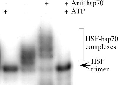
Figure 1.
Demonstration of HSF–hsp70 complexes. An extract that displays the multiple HSF bands characteristic of cells grown in low glucose was used for a gel mobility shift DNA-binding assay. ATP was added to the two outer lanes, and antibody to hsp70 was added to the two right lanes. The ladder of bands above the HSF trimer was specifically supershifted by the antibody and was specifically eliminated by ATP.

