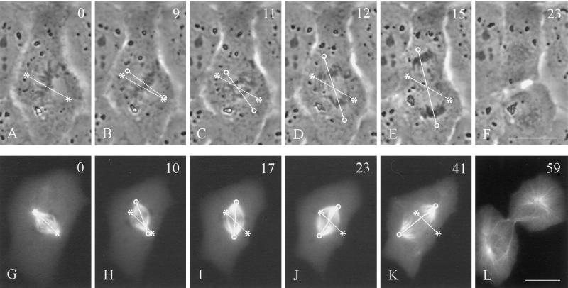
Figure 1.
Rotation of mitotic spindles in unmanipulated NRK cells. Time-lapse phase-contrast images (A–F) or microinjected rhodamine-tubulin images (G–L) were recorded in cells where the initial spindle orientation deviates from the long axis of the cell (A and G). Rotation of the spindle in the upper cell took place mostly in anaphase (C–E). Spindle rotation in the lower cell took place during metaphase (H–J). Both reached an orientation roughly along the longest axis of the cell (E and K). Time in minutes relative to metaphase is shown in the upper right corner. Asterisks and dotted lines indicate initial positions of the spindle poles and axis, respectively. Circles and solid lines indicate current positions of the spindle poles and axis, respectively. Bars, 20 μm.

