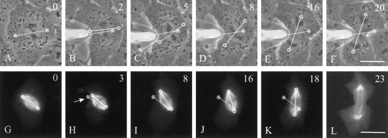
Figure 3.
Rotation of mitotic spindle in a cell manipulated to alter the orientation of the long axis. A blunted microneedle was gently placed against the membrane and moved forward to push the cortex (B). Soon after, deformation spindles began to rotate (C–D). By mid-anaphase, the spindle was aligned along the longest axis of the cell (E). The same approach was also used to deform the cortex of a cell microinjected with rhodamine-labeled tubulin (G–L), which had a shape similar to that for the cell in A–F. The position of the needle is shown with an arrow (H). The spindle rotated and the cell proceeded through anaphase and telophase, with no apparent damage to the spindle apparatus. Time relative to metaphase is shown in the upper right corner. Asterisks and dotted lines indicate initial positions of the spindle poles and axis, respectively. Circles and solid lines indicate current positions of the spindle poles and axis, respectively. Bars, 20 μm.

