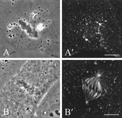
Figure 8.
The organization of dynein (A′) and microtubules (B′) in NRK cells microinjected with the 70.1 monoclonal antibody against dynein intermediate chain. Stacks of optical sections were deconvolved and used for reconstruction of the 90° view. Dynein staining revealed no localization along astral microtubules, although some staining is visible at the spindle poles (A and A′, arrows). The injection caused no apparent disruption of the spindle, as shown by immunofluorescence of tubulin in a separate cell (B and B′). Bars, 10 μm.

