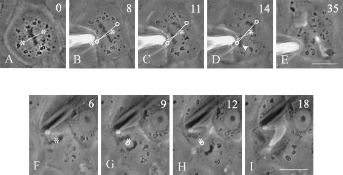
Figure 9.
Inhibition of spindle rotation and migration by anti-dynein antibodies. Two NRK cells were injected with 70.1 antibodies against the dynein intermediate chain and deformed with a microneedle as in Figures 3 and 4. In the first cell (A–E), the needle created a highly asymmetric cell shape. Throughout metaphase and anaphase the spindle remained close to its original orientation (B–D), such that one set of chromosomes bumped into the needle (D, arrowhead). A cleavage furrow developed along the longest axis of the cell between separating chromosomes. Asterisks and dotted lines indicate the initial positions of the spindle poles and axis, respectively. Circles and solid lines indicate current positions of the spindle poles and axis, respectively. In the second cell (F–I), the needle was used to create a constriction near one end of the cell, such that the cortex became much closer to one of the spindle poles. No repositioning of the spindle was detected. A cleavage furrow developed between separating chromosomes, creating two daughter cells of unequal sizes. Time relative to metaphase is shown in the upper right corner. Bars, 20 μm.

