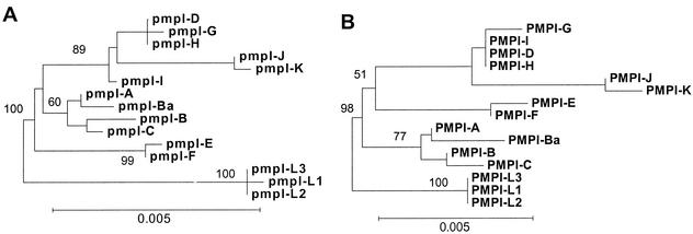
FIG. 2.
Evolution of pmpI shows weak segregation of serovars compatible with disease groups. Neighbor-joining trees based on the nucleotide (A) and inferred amino acid (B) sequences of pmpI are shown. Bootstrap values are shown only at the nodes separating major clades and only when they exceeded 50%. The scale bar equals a corrected sequence divergence of 0.005, which is the same as the scale used in Fig. 4 and 6. However, due to lesser divergence in pmpI, the branches and scale bar here are lengthened in order to plainly illustrate the branching patterns.

